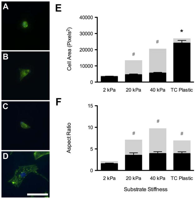
Figure 5. Effect of Cytochalasin-D on Cell Morphology.
Addition of the actin polymerization inhibitor, cytochalasin–D, caused the cells to adopt a more rounded morphology on 2 kPa (A), 20 kPa (B), and 40 kPa (C) gels and tissue culture plastic (D), and disrupted intracellular actin filaments (A–D, Green = F-actin, Blue = nuclei). Disrupting these actin filaments with cytochalasin–D resulted in significantly reduced cellular area for cells on 20 and 40 kPa gels compared to growth media values, but had no effect on ASCs on tissue culture plastic (E; black columns; cytochalasin–D media; grey columns: normal growth media). More importantly, however, it caused a significant reduction in cell aspect ratio for ASCs on 20 and 40 kPa gels and those cultured on standard tissue culture plastic compared to values in growth media (F, black columns; cytochalasin–D media; grey columns: normal growth media). There were no significant differences in aspect ratio between substrates in cytochalasin media. Scale bar = 50 μm. * indicates p < 0.001 compared to other groups in cytochalasin-supplemented media. # indicates p < 0.001 compared to same group in normal growth media.

