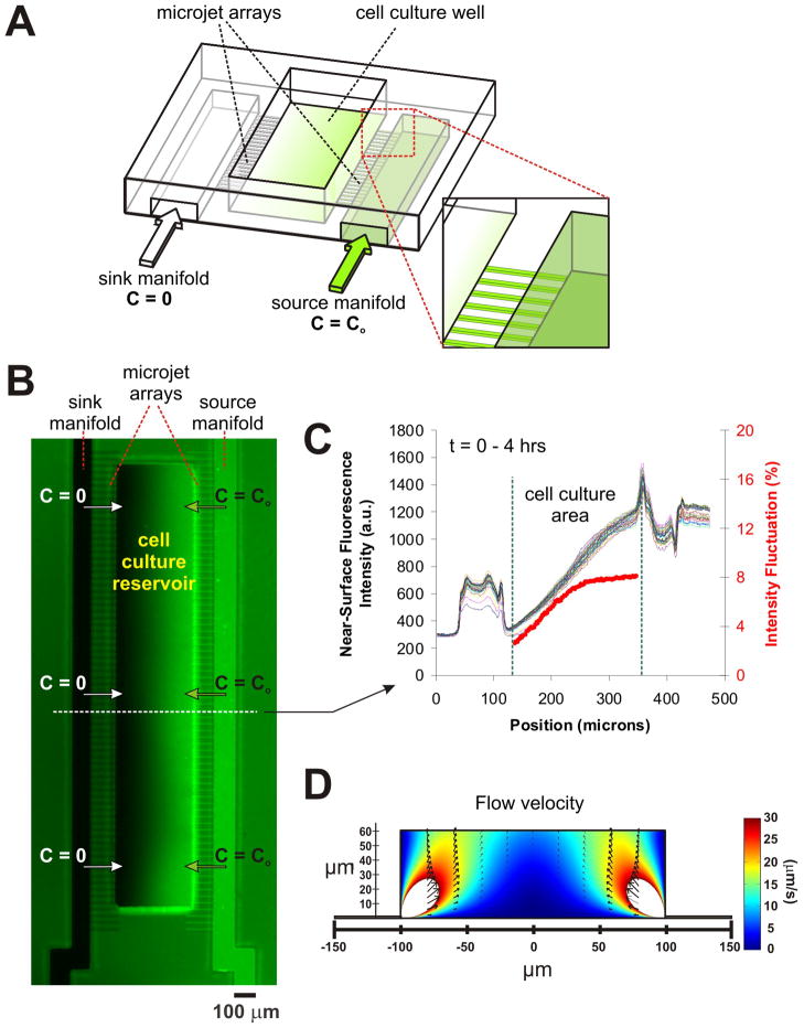
Fig. 1.
The Micro-jets Device: (A) Schematic of the device depicting a central open-surface reservoir (200 μm-wide, 66 μm-deep) that is fed laterally by two microchannels termed “sink manifold” and “source manifold” (each 100 μm-wide), which eject material into the reservoir through an array of small orifices called “micro-jets” (each approx. 10 μm × 2.5 μm cross section); (B) Pseudo-color image showing a representative, 4 hr-long surface gradient profile of fluorescein after the micro-jets are pressurized. The right microchannel (“source manifold”) was filled with 45 mM Orange-G and 1 mM fluorescein, while the left microchannel (“sink manifold”) and the cell culture reservoir were initially filled with 45 mM Orange-G only. (C) Line-scan measurements of fluorescence intensity across the device over time at the 10 pixel-wide line drawn in (A). The red line depicts the intensity fluctuations (defined as the standard deviation of all the fluorescence values over time observed for any given position of the channel relative to the time-average fluorescence at that position; the first four time points were not included for averaging because the gradient was still not in steady state). As shown, the gradient remained stable (within 3–8%) for 4 hours. (D) Fluid dynamic simulations plotting the flow velocities in a 60 μm high, 200 μm wide cell-culture reservoir. The arrows show that the flow is mostly directed upwards towards the air-fluid interface.

