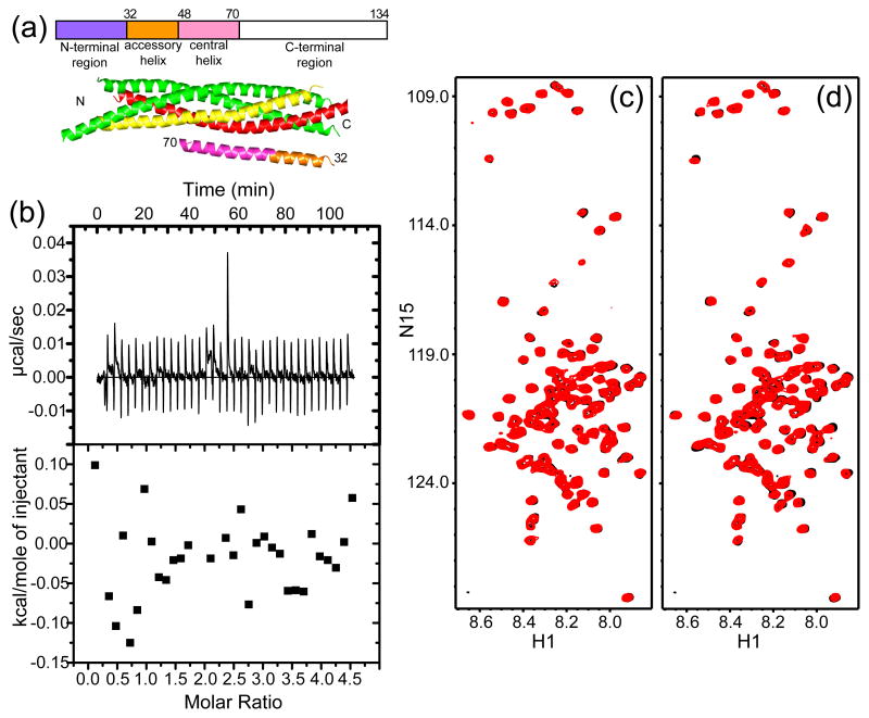
Figure 1.
Complexin-I does not bind to the synaptotagmin-1 C2AB fragment. (a) Domain diagram of complexin-I with residue numbers above, and ribbon diagram of the Cpx26-83/SNARE complex37 with SNAP-25 in green, syntaxin-1 in yellow, synaptobrevin in red and Cpx26-83 in orange (accessory helix) and pink (central helix). The N- and C-termini of the region of Cpx26-83 that was observable are labeled with residue numbers, and the N- and C-termini of the SNAREs are indicated by N and C, respectively. (b) ITC analysis of complexin-I binding to the C2AB fragment in 1 mM Ca2+. Complexin-I (150 μM) was titrated into the C2AB fragment (10 μM). (c,d) 1H-15N HSQC spectra of 8 μM 15N-labeled complexin-I in the absence (black) and presence (red) of 10 μM C2AB fragment and 1 mM EDTA (c) or 1 mM Ca2+ (d).

