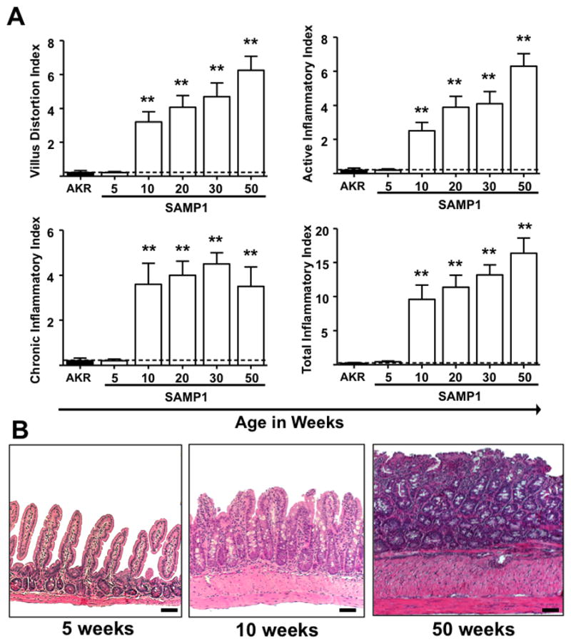
Figure 1. Time course and histological features of ileitis in SAMP1 mice.

Inflammatory indices (Villus distortion, active, chronic and total) were assessed by a pathologist (PJ) in a blinded fashion using a previously described scoring system, from 5- to 50-weeks-of-age. Normal villus, crypt and muscularis architecture was evident at 5-weeks-of-age. At 10-weeks-of age, transmural mixed inflammatory infiltrates; goblet and crypt hyperplasia and hypertrophy of the muscularis propria were present. Further progression of transmural infiltrates, villus distortion and muscularis hypertrophy were observed in 20- to 50-week-old mice with additional crypt elongation and prominent goblet cell hyperplasia. Scale bars represent 100μm. Data expressed as mean ± SEM, **P<.01 vs. total mean ± SEM for age-matched AKR controls from 5- to 50-weeks-of-age (n=5–9/age group).
