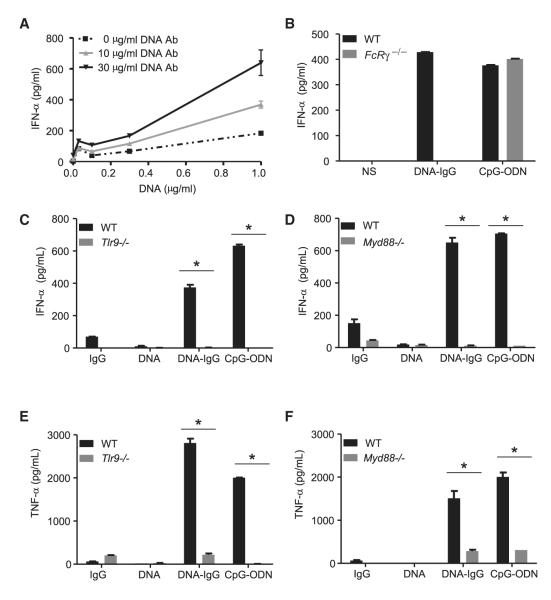
Figure 1. DNA-IgG-Mediated Production of IFN-α Requires Both FcγR and TLR9.
(A) Bone-marrow-derived pDCs from WT mice were stimulated with increasing concentrations of CG50 plasmid DNA (DNA) in presence of different concentrations of the DNA antibody E11 (DNA Ab). Production of IFN-α was measured by ELISA after 24 hr.
(B) Bone-marrow-derived pDCs from WT and Fcer1g (FcRγ −/−) mice were unstimulated (NS) or stimulated with DNA-IC formed by combining CG50 plasmid DNA and DNA antibody E11 (DNA-IgG). We used 5 μM of ODN1585 (CpG-ODN) as positive control. Secretion of IFN-α was measured by ELISA 24 hr after pDC stimulation. Data is representative of three independent experiments.
(C–F) Fetal liver-derived pDCs from WT, Tlr9−/− (C and E) or Myd88−/− (D and F) mice were stimulated with DNA antibody E11 (IgG), CG50 plasmid DNA (DNA), or DNA-IgG. ODN1585 (CpG-ODN) was used as a positive control. IFN-α and TNF-α in supernatants were measured by ELISA 24 hr after pDC stimulation. Data are presented as mean ± SD of three independent experiments. (*p < 0.05).

