Figure 2.

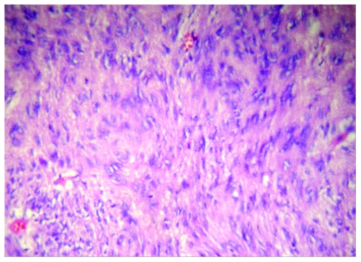
Composition of the tumor. Light microscopy revealed that the tumor primarily consisted of spindle cells (hematoxylin and eosin staining; magnification, ×200).

Composition of the tumor. Light microscopy revealed that the tumor primarily consisted of spindle cells (hematoxylin and eosin staining; magnification, ×200).