
Figure 2.
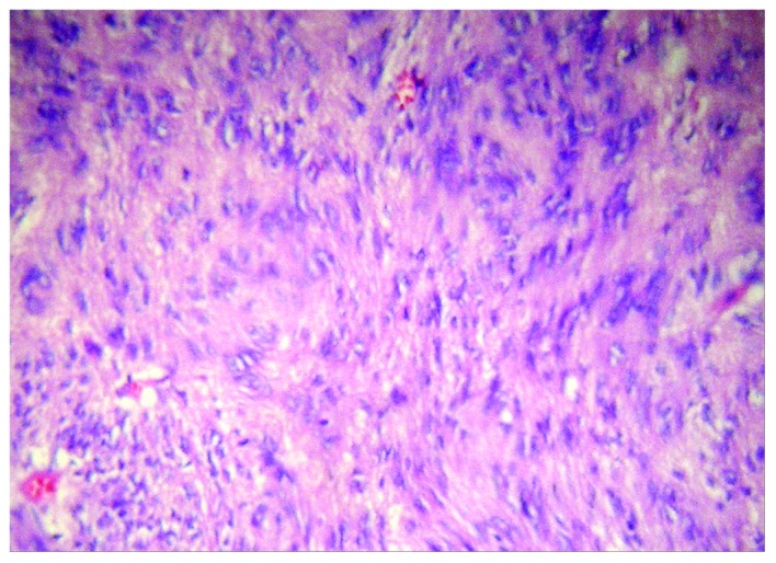
Composition of the tumor. Light microscopy revealed that the tumor primarily consisted of spindle cells (hematoxylin and eosin staining; magnification, ×200).
Official websites use .gov
A
.gov website belongs to an official
government organization in the United States.
Secure .gov websites use HTTPS
A lock (
) or https:// means you've safely
connected to the .gov website. Share sensitive
information only on official, secure websites.
Composition of the tumor. Light microscopy revealed that the tumor primarily consisted of spindle cells (hematoxylin and eosin staining; magnification, ×200).