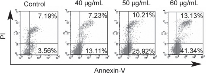
Figure 1.

Effects of propranolol on the apoptosis of IHECs. IHECs were left untreated or exposed to 40, 50 or 60 μg/ml propranolol for 24 h and then stained with Annexin-V and PI. Apoptosis was analyzed with flow cytometry. Cell populations in the upper (late apoptosis) and lower (early apoptosis) right quadrants were designated as apoptotic cells. Representative dot plots of three separate experiments with similar results are shown. IHECs, infantile hemangioma endothelial cells; PI, propidium iodide.
