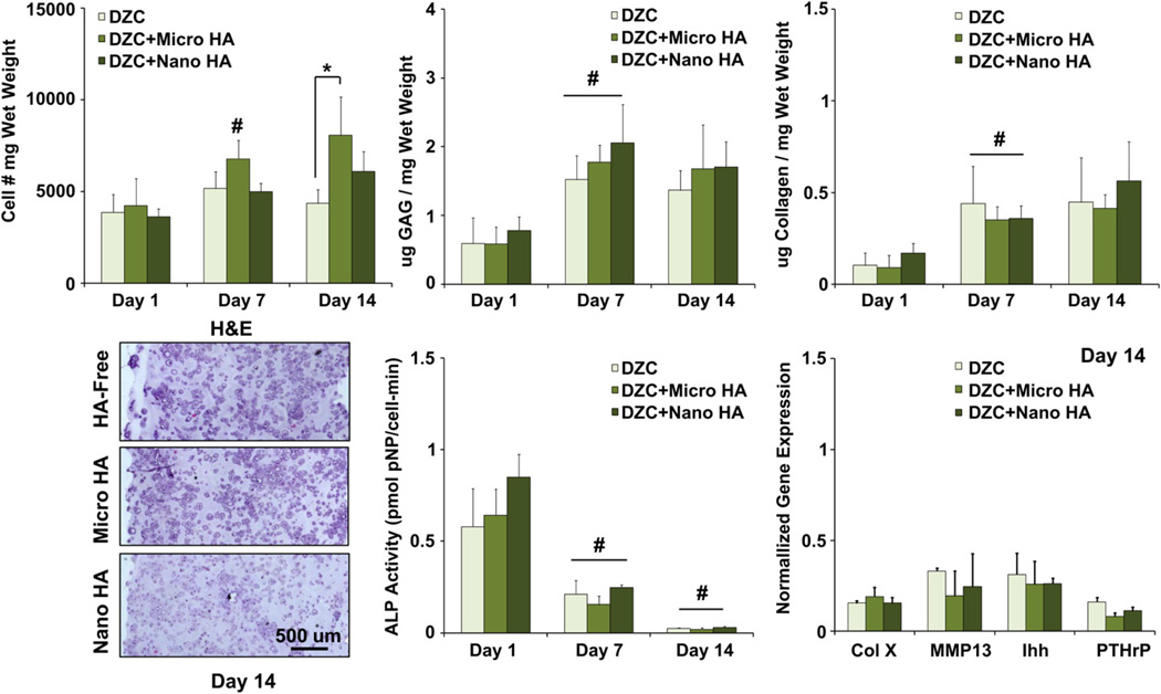
Fig. 2.
Effect of HA presence and size on deep zone chondrocytes. A higher cell number was measured at day 14 for the micro-HA group as compared to HA-free control (*p < 0.05, n = 5). Both cells and matrix are uniformly distributed throughout the scaffolds (H&E, 10x, bar = 500 µm, Day 14, n = 2), and GAG as well as collagen content increased for all groups over the first week of culture (#p < 0.05, n = 5). Cell ALP activity decreased over time for all groups (#p < 0.05, n = 5), with no change detected in the expression of hypertrophic markers due to either the presence or size of HA particles (n = 3).

