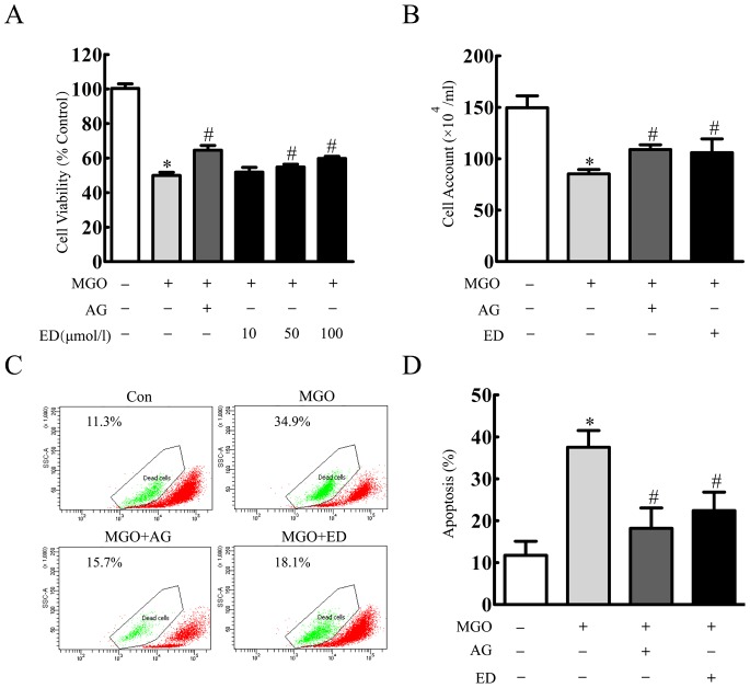
Figure 2. Edaravone protected MGO-induced cell injury in the cultured HBMEC.
(A) HBMEC were incubated with edaravone (10 µmol/l, 50 µmol/l, 100 µmol/l), aminoguanidine (1 mmol/l) for 20 min before MGO (2 mmol/l) treatment. After 24 h MGO treatment, cell viability was determined by MTT assay. (B) Cell counts were assessed by trypan blue exclusion. (C) HBMEC were incubated with edaravone (100 µmol/l), aminoguanidine (1 mmol/l) 20 min before MGO (2 mmol/l) treatment. After 24 h MGO treatment, cell apoptosis was detected by rhodamine 123 staining and flow cytometer. *p<0.05 versus control group. #p<0.05 versus MGO group. n = 3 repeats. AG represented aminoguanidine. ED represented edaravone.

