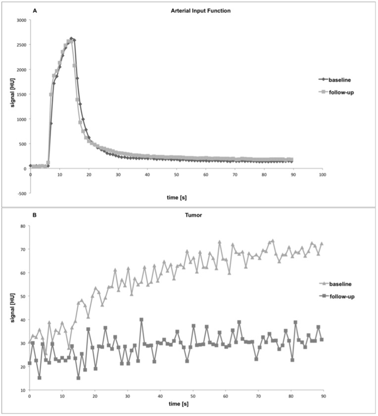
Figure 2. Representative arterial input functions (A) and tumor signal enhancement curves (B) at baseline and follow-up. Signal intensity [HU, Hounsfield Units] (y-axis) is displayed over time [s] (x-axis).
Note the almost identical arterial input functions for baseline and follow-up which can be considered a marker of good reproducibility of individual perfusion scans. After anti-angiogenic therapy, a decline in tumor signal enhancement due to reduced tumor perfusion was observed.

