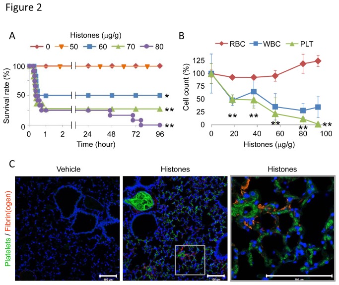
Figure 2. Extracellular histones cause fatal thromboembolism in mice.
(A) The lethal effect of extracellular histones. Mice were intravenously injected with histones (0-80 µg/g, n = 7-12 per group), and survival was analyzed. (B) Histone-induced thrombocytopenia. Numbers of platelets (PLT), red blood cells (RBC), and white blood cells (WBC) in blood 10 min after infusion with histones (0-95 µg/g, n = 3-7 per group, mean ± S.D.) are shown. Data are presented as percentage of the vehicle group (0 µg/g histones). (C) Distribution of DyLight488-labeled platelets and Alexa-Fluor 594-labeled fibrin(ogen) in lung tissue 10 min after infusion with vehicle or 75 µg/g histones. Nuclei were stained with DAPI. Representative images of n = 4. Scale bar = 100 µm. * P < 0.05 and ** P < 0.01 compared with the vehicle group.

