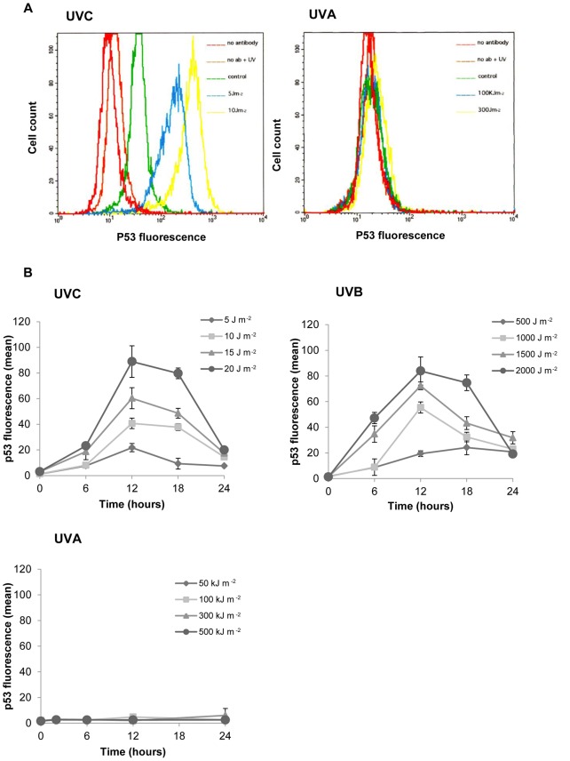
Figure 2. Kinetics and dose dependence of p53 accumulation as determined by fluorescence-activated cell sorting.
Figure 2A. Representative flow cytograms of fibroblasts, stained for p53 with a fluorescein isothiocyanate-conjugated antibody. Cells were exposed to ultraviolet radiation in the exponential phase of growth and assayed 6 hours post insult. Figure 2B. P53 accumulation in G1 irradiated fibroblasts in response to ultraviolet radiation. Each data point represents the mean ± S.E.M. for at least 3 independent experiments. S.E.M = Standard error of the mean.

