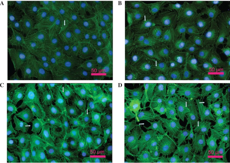
Figure 2.
Effect of lipopolysaccharide (LPS) and high glucose on the actin cytoskeleton in human pulmonary microvascular endothelial cells (PMVECs). Cells were incubated with normal (5.5 mM) or high (33 mM) D-glucose concentrations for 5 days in medium with 2% serum (to maintain the cells in the quiescent state) and then cells were incubated with LPS (10 μg/ml) for 12 h. Cells were stained for F-actin and visualized under a fluorescence microscope as described in Materials and methods. Scale bars, 50 μm. (A) Normal glucose group; (B) high glucose group; (C) normal glucose + LPS group; (D) high glucose + LPS group.

