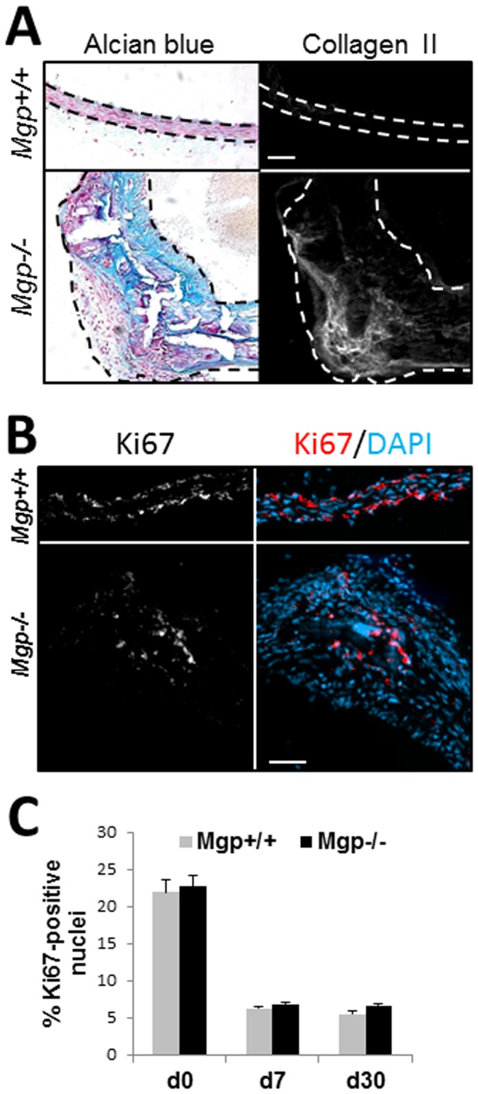
Figure 1. Cartilaginous metaplasia and cell proliferation in the Mgp-/- aorta.

A, Detection of cartilage matrix using Alcian blue staining for GAG deposition and immunostaining for Collagen type II (Collagen II) protein on adjacent sections of aorta from 4.5 week old wild-type (Mgp+/+) and Mgp-/- animals. Scale = 50 µm. Dashed lines denote internal and external elastic lamina. B, Representative immunostaining for cell proliferation marker, Ki67 (white, red) [nuclei counterstained with DAPI (blue)] in 30 day old Mgp+/+ and Mgp-/- aortae. Scale = 50 µm. C, Quantitation of the percentage of Ki67-positive nuclei compared to total nuclei in Mgp+/+ and Mgp-/- aortic tissue from animals at birth (d0, N=3), at 7 days old (d7, N=4), and at 30 days old (d30, N=4). Four sections per animal were analyzed.
