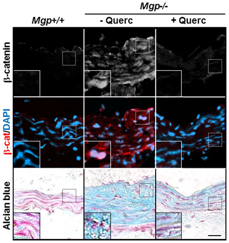
Figure 6. Quercetin blocks accumulation and nuclear localization of β-catenin protein in Mgp-/- aortae.
Immunostaining for β-catenin (white, red) [nuclei counterstained with DAPI (blue)] and Alcian blue staining for GAG deposition on adjacent sections of aortae from Mgp+/+, untreated Mgp-/-, and quercetin-treated Mgp-/- animals shows that β-catenin protein is detected only in untreated Mgp-/- arterial tissue. Scale = 30 µm. Inset panels show higher magnification of representative nuclei, demonstrating co-localization of β-catenin with DAPI in the Mgp-/- aorta in rounded chondrocyte-like cells surrounded by cartilaginous GAG-rich matrix.

