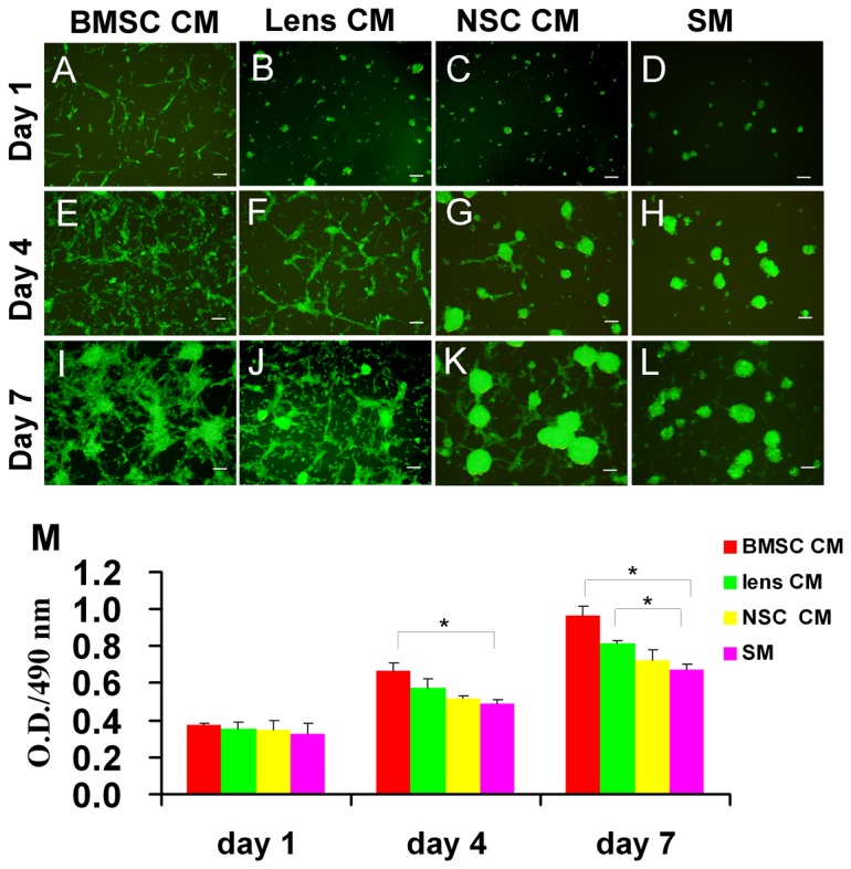
Figure 1. Morphology and proliferation capacity of CM-treated RPCs.

In the proliferation conditions, the RPCs were cultured in BMSC, lens and NSC CM, and morphology images were taken on days 1 (A, B, C, D), 4 (E, F, G, H) and 7 (I, J, K, L). In the presence of BMSC CM, the cells attached to the surface of the flask and extended short processes by the first day; with time, most cells exhibited two or more long processes and formed an intercellular network by day 7 (A, E, I). With lens CM, the cells were adherent, and short processes extending from a few cells were observed by day 1; with time, most cells extended processes, but there were fewer adherent cells than in the BMSC CM condition (B, F, J). In the presence of NSC CM (C, G, K) or SM, (D, H, L), most RPCs grew as spherical clusters which adhered to the flask or floated in the culture medium. The spherical cluster size in NSC CM was larger than that in SM. The expansion potential of the RPC cultures was assessed using CCK-8 analysis. The cells exhibited an obvious increase in expansion potential in CM cultures, especially in the BMSC CM, than in SM cultures (M). Scale bars: 100 µm.
