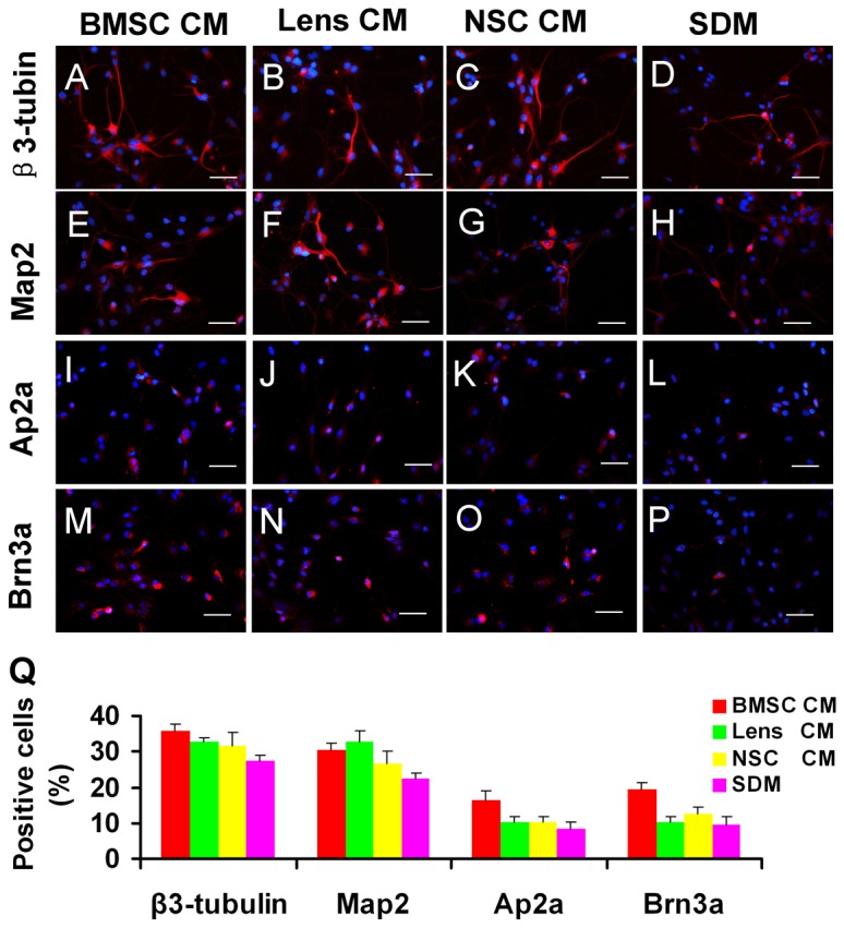
Figure 6. Potential of RPC differentiation towards neurons after exposure to CM.

After RPCs were cultured in the differentiation condition for 7 days, the cells were immunolabelled for anti-β3-tubulin (A-D), -Map2 (E-H), -AP2α (I-L) and -Brn3a (M-P). The proportion of β3-tubulin, AP2α and Brn3a-positive cells was highest in BMSC CM-treated RPCs (Q). The percentage of MAP2-immunoreactive cells was significantly higher in lens and BMSC CM cultures (Q). The quantification of immunoreactive cells was performed as described in Figure 3-I. *P<0.05. Scale bars: 50 µm.
