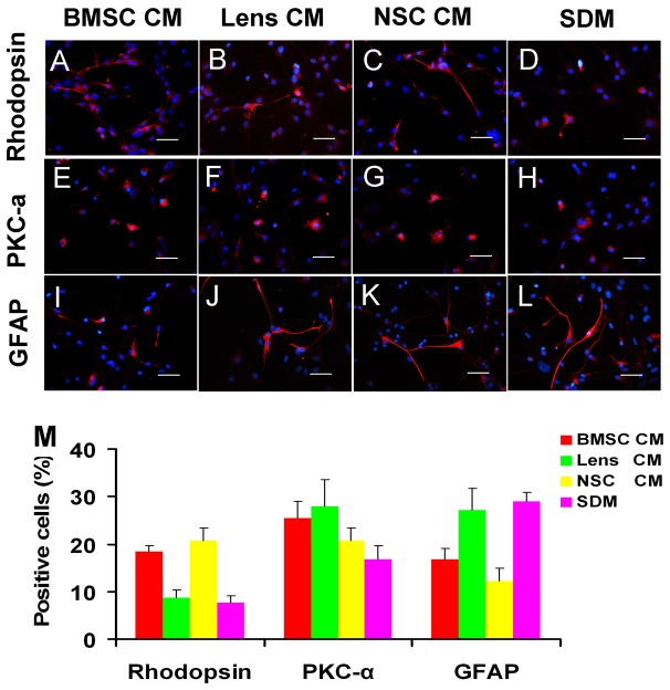
Figure 7. Potential of RPC differentiation towards neuronal and glial cells after exposure to CM.
After RPCs were cultured in differentiation medium for seven days, the cells were fixed and immunostained with antibodies against rhodopsin (A-D), PKC-α (E-H) and GFAP (I-L). The percentages of rhodopsin-positive cells were higher in NSC and BMSC CM-treated RPC cultures than in other groups, and PKC-α immunoreactive cells were detected more in CM treated RPCs. However, the ratio of GFAP-positive cells was decreased in the BMSC and NSC CM-treated cultures compared with the controls (M). Quantification of immunoreactive cells was performed as described in Figure 3-I. *P<0.05. Scale bars: 50 µm.

