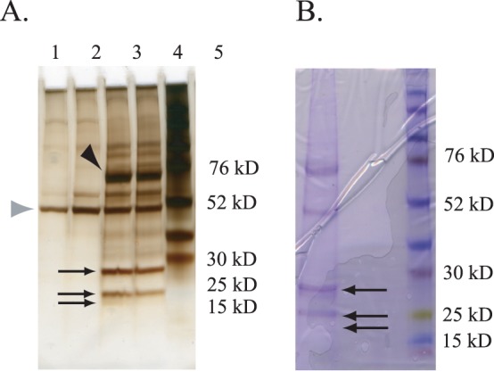
Figure 4. Purification of three Morgue-associated proteins.

A. Silver staining of an analytical SDS polyacrylamide gel separating proteins from whole fly extracts that associate with Morgue. Protein bands corresponding to Morgue-3xFLAG (∼60 kD) (black arrowhead) as well as the anti-FLAG Ig heavy chain (∼50 kD) are indicated (gray arrowhead). Lanes 1–4 correspond to material purified via the anti-FLAG resin from the following flies: Lane 1: P[da-Gal4]; Lane 2: P[UAS-Morgue3xFlag]; Lane 3: P[da-Gal4], P[UAS-3xFlag:Morgue]; Lane 4: P[da-Gal4], P[UAS-Morgue3xFlag]; Lane 5: Protein molecular weight markers. The three major bands corresponding to Morgue-associated proteins purified are indicated (arrows) and correspond to polypeptides migrating between 28 kD and 20 kD. B. Coomassie Brilliant Blue staining of a preparatory SDS polyacrylamide gel separating Morgue-associated proteins from extracts of P[da-Gal4], P[UAS-Morgue:3xFlag] adult flies purified via anti-FLAG resin. Three major bands (arrows) were excised from the gel and the corresponding polypeptides analyzed via mass spectrometry. Lane on right corresponds to protein molecular weight markers.
