
Figure 4. Disulfide-linked GARP/TGF-β1 complexes are released in the supernatant of T cells, but not 293 cells.
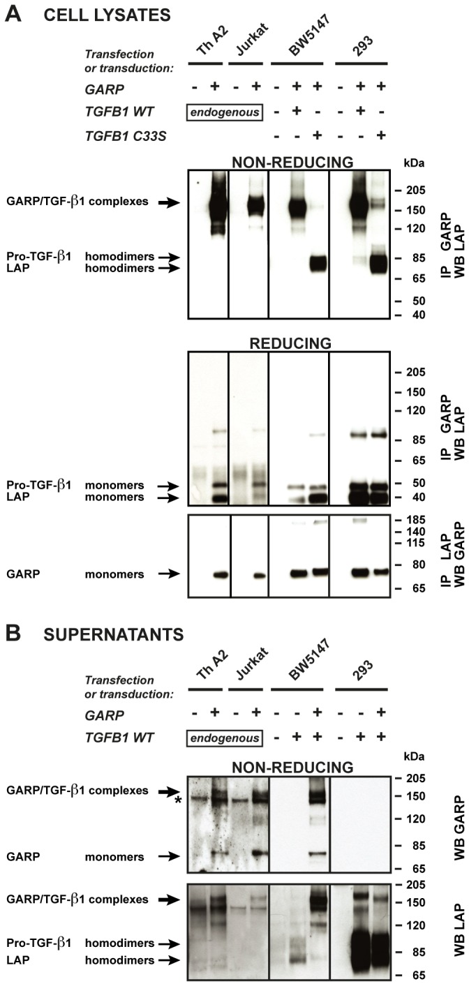
A. Cells described in Figure 2 were lysed and immunoprecipitated (IP) with anti-GARP or anti-LAP antibodies. IP products were submitted to SDS-PAGE under non-reducing or reducing conditions, followed by WB with anti-LAP antibodies (top and middle panels), or anti-GARP antibodies (bottom panels). Pro-TGF-β1 and LAP homodimers in the top panels are not clearly resolved, but can be distinguished better with longer migrations or higher concentrations of polyacrylamide. The +/- 85-90 kDa bands that appear in the middle panel correspond to non-specific bands, or to incompletely reduced pro-TGF-β1. B. Cells (2x10⁶/ml for murine and human T cells, 2.5x10⁵/ml for transfected 293 cells) were incubated in serum free medium during 24 hours. Different cell concentrations were used to adjust for the different amounts of secreted TGF-β1 (see Figure 2). Human Th A2 and Jurkat cells were stimulated with anti-CD3/CD28 antibodies to increase secretion. Supernatants (0.5-10 µl) were analyzed by WB under non-reducing conditions with anti-GARP and anti-LAP antibodies. * Band that also appears when the secondary anti-IgG2b-HRP antibody is used alone (without anti-GARP antibody), due to cross reactivity against the anti-CD3/CD28 antibodies used for T cell stimulation.