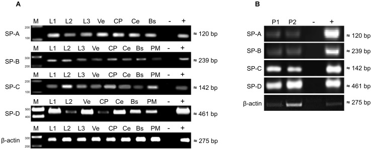
Figure 1. RT-PCR analysis of human brain.
Detection of SP-A, SP-B, SP-C and SP-D in A) tissue specimens of brainstem (BS), cerebellum (Ce), choroid plexus (CP), subventricular cortex (Ve), pia mater (PM) and cerebrospinal fluid (L1, L2, L3) and B) in pineal gland (P1, P2). DEPC-H2O served as negative control (−), RNA-extract from lung tissue was used as positive control (+), molecular size was estimated using a molecular marker (M).

