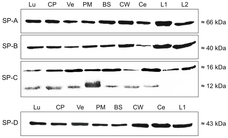
Figure 2. Western blot analysis of surfactant proteins.
Detection of SP-A, SP-B, SP-C and SP-D after SDS gel electrophoresis in protein isolates of brainstem (BS), cerebellum (Ce), choroid plexus (CP) the circle of Willis (CW), subventricular cortex (Ve), leptomeninx (PM) and cerebrospinal fluid (L1, L2), Lung (Lu) served as positive control.

