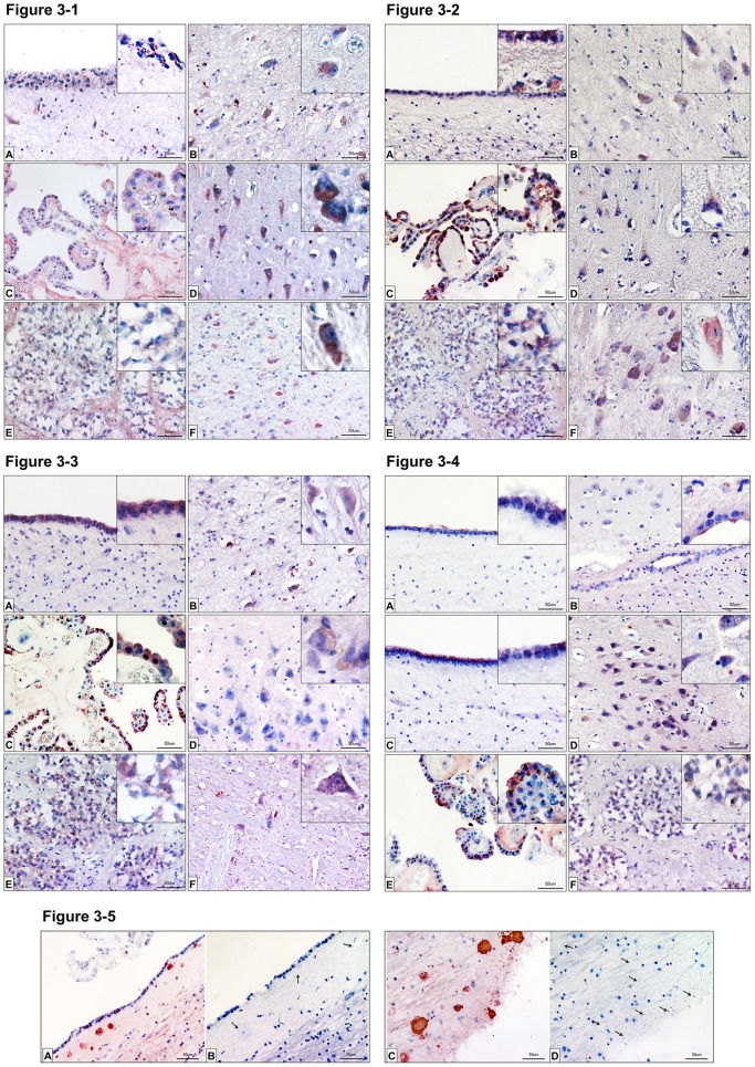
Figure 3. Immunhistochemistry of surfactant proteins A, B, C and D.
Figure 3-1: Immunohistochemistry of SP-A: Detection of surfactant protein A in the CNS by means of immunohistochemistry. Sections from the ventricles [A], the hilum region of the hippocampus [B], choroid plexus [C], the dentate gyrus [D], pineal gland [E] and medulla oblongata [F] were analyzed. Red staining indicates SP-A occurrence. Insets in the figures show magnifications for the respective tissue. Control sections (secondary antibody only) were negative (unstained) for each tissue. Scale bars: 50 µm. Figure 3-2: Immunohistochemistry of SP-B: Detection of surfactant protein B in the CNS by means of immunohistochemistry. Sections from the ventricles [A], the hilum region of the hippocampus [B], choroid plexus [C], the dentate gyrus [D], pineal gland [E] and medulla oblongata [F] were analyzed. Red staining indicates SP-B occurrence. Insets in the figures show magnifications for the respective tissue. Control sections (secondary antibody only) were negative (unstained) for each tissue. Scale bars: 50 µm. Figure 3-3: Immunohistochemistry of SP-C: Detection of surfactant protein C in the CNS by means of immunohistochemistry. Sections from the ventricles [A], the hilum region of the hippocampus [B], choroid plexus [C] the dentate gyrus [D], pineal gland [E] and medulla oblongata [F] were analyzed. Red staining indicates SP-C occurrence. Insets in the figures show magnifications for the respective tissue. Control sections (secondary antibody only) were negative (unstained) for each tissue. Scale bars: 50 µm. Figure 3-4: Immunohistochemistry of SP-D: Detection of surfactant protein D in the CNS by means of immunohistochemistry. Sections from the ventricles [A, C], the hilum region of the hippocampus [B] and near the dentate gyrus [D], choroid plexus [E], and pineal gland [F] were analyzed. Red staining indicates SP-D occurrence. Inserts in the figures show magnifications for the respective tissue. Control sections (secondary antibody only) were negative (unstained) for each tissue. Scale bars: 50 µm. Figure 3-5: Immunohistochemistry of SP-C plaques: Detection of surfactant protein C in the CNS by means of immunohistochemistry. Sections from fossa rhomboidea stained [A–C], and unstained [C–D] for SP-C were analyzed. Red staining indicates SP-C occurrence. Insets in the figures show magnifications for the respective tissue. Control sections (secondary antibody only) were negative (unstained) for each tissue. Arrows indicate the possible plaque sites, neuromelanin granules and possibly degenerated neurons. Scale bars: 50 µm.

