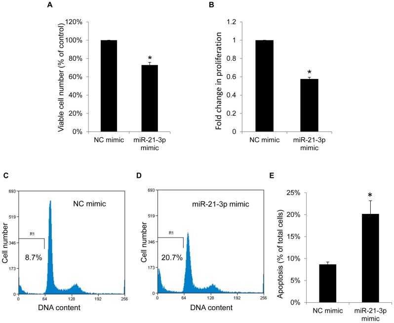
Figure 6. MicroRNA-21-3p suppresses growth and induces apoptosis in HepG2 cells.
(A) After 50 nM of miR-21-3p mimic or negative-control mimics were transfected for 48 h, viable HepG2 cells were quantified using a Trypan blue dye exclusion assay. (B) After transfecting 50 nM of miR-21-3p mimics or negative-control mimics into HepG2 cells for 24 h and incubating for an additional 24 h for BrdU incorporation, the cellular proliferation was measured using the BrdU incorporation assay kit and expressed as fold change. (C, D and E) After 50 nM of miR-21-3p mimics or negative-control mimics were transfected for 72 h, apoptosis was detected by measuring the sub-G1 population using flow cytometry with propidium iodide staining. Data are presented as the mean ± standard deviation of 3 independent experiments. “*” indicates a significant difference with P<0.05.

