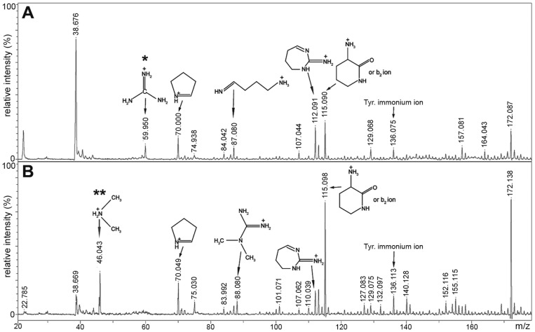
Figure 2. Detection of diagnostic arginine and asymmetric dimethylarginine ions.
Zoomed view from region m/z 20–175 of MALDI-TOF/TOF analysis of (A) m/z 623.2, corresponding to unmodified peptide 251-GGGRGGY-257 from recombinant hnRNP A2 and (B) m/z 651.3, corresponding to peptide 251-GGGRGGY-257 with a dimethylarginine modification from HPLC-purified rat brain hnRNP A2. Structures representing diagnostic ions of arginine* and dimethylarginine** are labeled as previously described [30]. See Supporting Information Fig. S4 for full spectrum.

