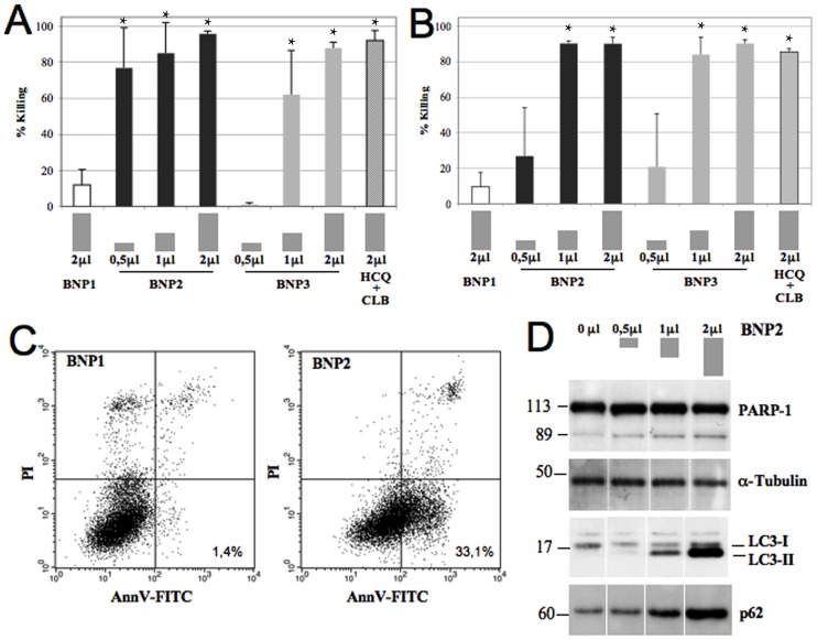
Figure 2. In vitro characterization of the cytotoxic effect of BNP2.
BJAB (A) and Raji (B) cells were incubated with 0.5, 1 and 2 μL of BNPs or HCQ+CLB for 48 hours at 37°C and residual viable cells were measured. Data are expressed as mean ± SD. *: p<0.01 vs BNP1. C) BJAB cells wer incubated with 1 μL of BNPs for only 16 hours at 37°C and apoptotic cells were analyzed using AnnexinV/PI test. D) Western blot analysis of activated PARP-1, LC3 and p62 from cell lysates obtained from BJAB cells incubated with 0, 0.5, 1 and 2 μL of BNP2.

