
Figure 4. Immunohistochemistry for sFlt-1 protein expression in chorionic villus.
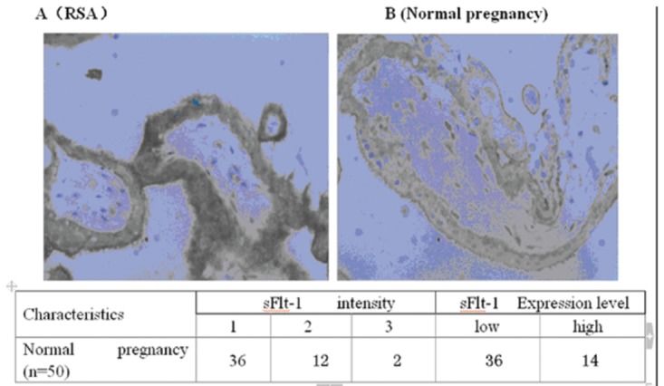
sFlt-1 protein was expressed in the chorionic villus located in the cytoplasm (Original magnification ×600. sFlt-1 intensity by immunohistochemistry staining was scored as following: 1=weak staining; 2=mild staining and 3=dark staining. *Based on the score of sFlt-1 intensity, cases were classified into sFlt-1 low-expression (low) for those cases in which of sFlt-1 intensity scores were ≤1 and sFlt-1 high-expression (high) for those cases in which of sFlt-1 intensity scores were ≥2. Scores were generated and analyzed by LTQ-Orbitrap mass spectrometry. We found that the chorionic villus of RSA women that subsequently miscarried showed a high expression of sFlt-1 (n =26) (A), whereas this tissue showed a low expression of sFlt-1 during early pregnancy in women with normal pregnancies (n =18) (B).