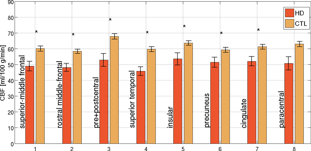
Figure 2. Region-of-Interest (ROI) analysis in regions found to be the most affected in HD.
While the normative resting CBF varies across the cortex, a similar degree of proportionate CBF reduction in HD was observed across all ROIs. The error bars represent standard error, and significant differences are denoted by asterisks (p < 0.05). In many ROIs, reductions in cortical CBF in HD subjects exceed the degree of concurrent cortical thinning in the same regions, suggesting that the two were not entirely associated. The error bars represent standard error, and significant differences are denoted by asterisks (p < 0.05).

