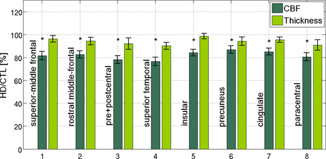
Figure 3. Regional associations and dissociations between CBF and cortical thickness reductions in HD.
CBF reductions were present across the cortical mantle, most prominently in the medial superior-frontal cortex, superior temporal gyrus and selectively in the occipital lobe. CBF reduction (red) and cortical thinning (orange) were associated (overlap shown in yellow) in the superior frontal, precentral, lateral occipital and posterior cingulate regions. On the other hand, spatial mismatches between the CBF and cortical thickness effects were evident in the insula, medial frontal and postcentral gyri as well as the lateral occipital lobe.

