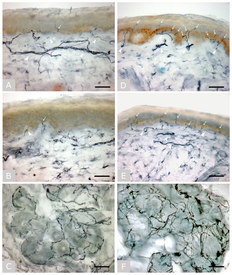
Figure 1. Skin biopsy.
Bright field immunohistochemical studies (polyclonal anti-protein gene product 9.5 antibody; Ultraclone, Wellow, Isle of Wight, UK) in sections of skin biopsy taken at the proximal thigh (A) and distal site of the leg (B) with a sweat gland (C) in patient no. 7, and at the proximal thigh (D) and distal site of the leg (E) with a sweat gland (F) in a healthy subject.
Arrows indicate intra-epidermal nerve fibers and arrowheads indicate dermal nerve bundles. The patient, with normal nerve conduction study findings and myopathy at needle electromyography, showed a severe depletion of intra-epidermal nerve fibers and a severe reduction in the density of dermal nerve bundles, whose staining was fragmented and weaker due to axonal degeneration. Sweat gland innervation was also markedly reduced. Original magnification 40x. Bar is 50 µm in all images.

