
Figure 2.
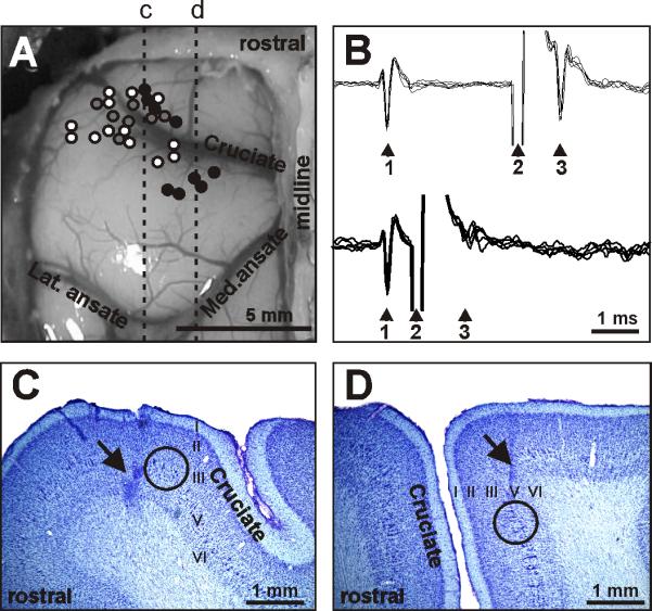
Location and identification of neurons. A: Areas of recording in the left motor cortex. Microelectrode entry points into the cortex were combined from all cats and are shown as circles on the photograph of cat 2 cortex: cat 1, 2, and 3 entry points are depicted by grey, white, and black circles, respectively. Positions of parasagittal sections, whose photomicrographs are shown in C and D are indicated by dotted lines c and d, respectively. B: A collision test determines whether a neuron's response to pyramidal tract stimulation is antidromic. Top: the neuron spontaneously discharges (arrowhead 1) and the pyramidal tract is stimulated approximately 3 ms later (arrowhead 2). The neuron responds with a latency of approximately 1 ms (arrowhead 3). Bottom: the neuron spontaneously discharges (arrowhead 1) and the pyramidal tract is stimulated about 0.5 ms later (arrowhead 2). The neuron does not respond (arrowhead 3) because at 0.5 ms the spontaneous spike was still en route to pyramidal tract, therefore causing the collision, or nullification, of the response. The test confirms that the neurons's response was antidromic, and therefore the neuron proves to be a pyramidal tract projecting neuron (PTN). C: Photomicrograph of a parasagittal section through the lateral pre-cruciate motor cortex (section c). An arrow points to a reference electrolytic lesion in the forelimb representation of the motor cortex. D: Photomicrograph of a parasagittal section through the medial motor cortex (section d). An arrow points to a reference electrolytic lesion in the hindlimb representation of the motor cortex. In C and D: Layers of the cortex are numbered. Groups of giant cells in layer V, which are characteristic for area 4γ and are visible throughout both the pre- and post-cruciate cortex, are encircled. Cresyl violet stain.