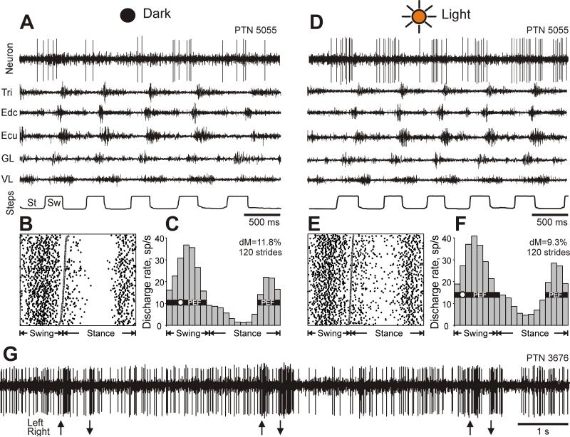
Figure 3.
A typical example of the activity of a neuron (PTN 5055) and selected right fore- and hindlimb muscles during locomotion in the darkness and light. A: Activity of the neuron and muscles during locomotion in the darkness. Tri, m. triceps brahii (elbow extensor); Edc, m. extensor digitorum communis (wrist and phalanges dorsal flexor); ECU, m. extensor carpi ulnaris (wrist dorsal flexor); GL, m. gastrocnemius lateralis (ankle extensor), VL, m. vastus lateralis (knee extensor). The bottom trace shows the stance (St) and swing (Sw) phases of the step cycle of the right forelimb that is contralateral to the recording site in the cortex. B, C: The activity of the same neuron during locomotion in the darkness is presented as a raster of 120 step cycles (B) and as histograms (C). The duration of step cycles is normalized to 100%. In the raster, the end of swing and the beginning of the stance in each cycle is indicated by an open triangle. In the histogram, the horizontal black bar shows the period of elevated firing (PEF) and the circle indicates the preferred phase as defined in the “Methods” section. The value of dM is stated. D-F: Activities of the same neuron and muscles during locomotion in illuminated room. G: Responses of a neuron to movements of an object in front of the cat (PTN 3676, located in the rostral cruciate sulcus). Arrows pointed up indicate movements from cat's right to left, arrows pointed down indicate movements from cat's left to right.

