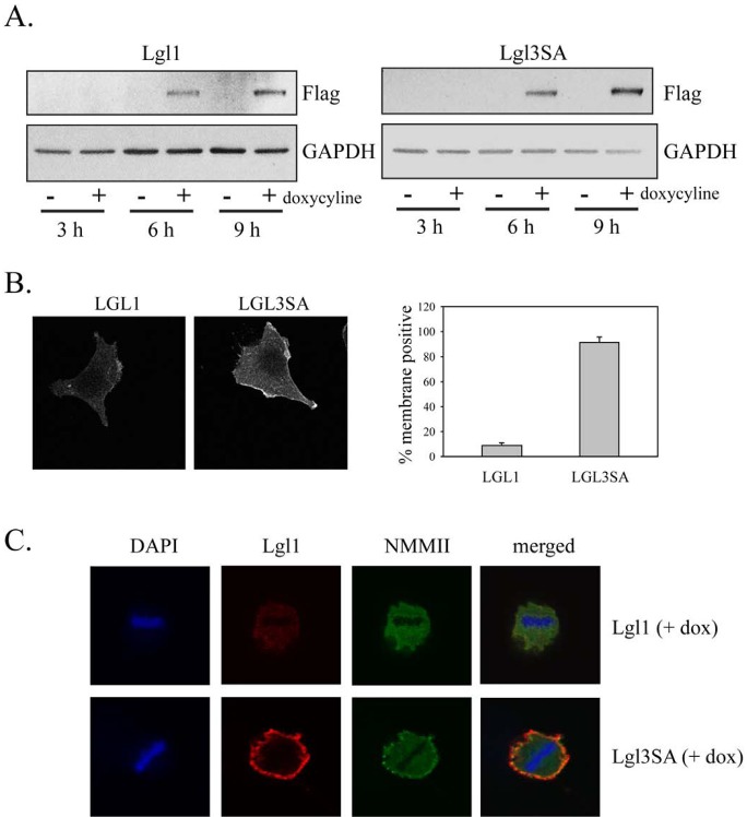
Figure 3. Non-phosphorylatable Lgl1 constitutively associates with the cell membrane in U87MG cells.
U87MG cells were transduced with lentiviral vectors expressing tet activator and tet-inducible Lgl or Lgl3SA. Cells were treated with 100 ng/ml doxycycline for the indicated periods of time and then analyzed by Western blotting with anti-Flag antibody. B. U87MG cells with inducible Lgl1 or Lgl3SA expression were treated with 100 ng/ml doxycycline for 4 days and then analyzed by immunofluorescence microscopy using anti-Flag antibody. Membrane localization was quantitated as describe in Figure 2C. C. U87MG cells with inducible Lgl1 or Lgl3SA expression were treated with 100 ng/ml doxycycline for 4 days and then analyzed by confocal immunofluorescence microscopy with antibodies to Flag epitope (red) and non-muscle myosin IIa (green). Cells were counterstained with DAPI. Examples of confocal images of mitotic cells expressing Lgl1 and Lgl3SA are shown.

