
Figure 7. Effects of PKCι on PriGO8A differentiation.
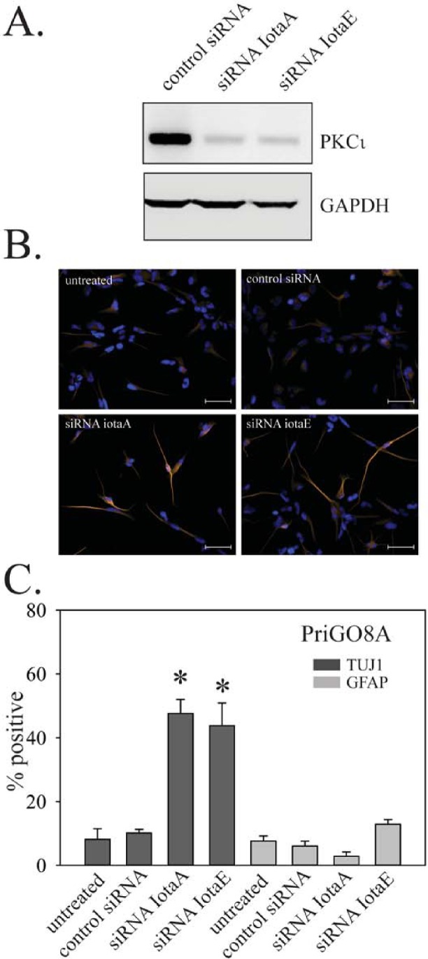
A. PriGO8A cells were transfected with control RNA duplex or two different RNA duplexes targeting PKCι. Three days later, total cell lysates were collected and analyzed for PKCι expression by Western blotting. B. Cells were transfected as in A. Seven days later cells were fixed and immunofluorescence with antibodies to TUJ1 and GFAP was performed. Representative images of TUJ1 immunofluorescence are shown. C. Quantitation of TUJ1 and GFAP immunofluorescence seven days after PKCι knockdown. Quantitation was performed as described in Material and Methods. Data are shown as the mean ± SE. * indicates a p value less than 0.05.