
Figure 8. Effects of Lgl3SA on PriGO8A differentiation.
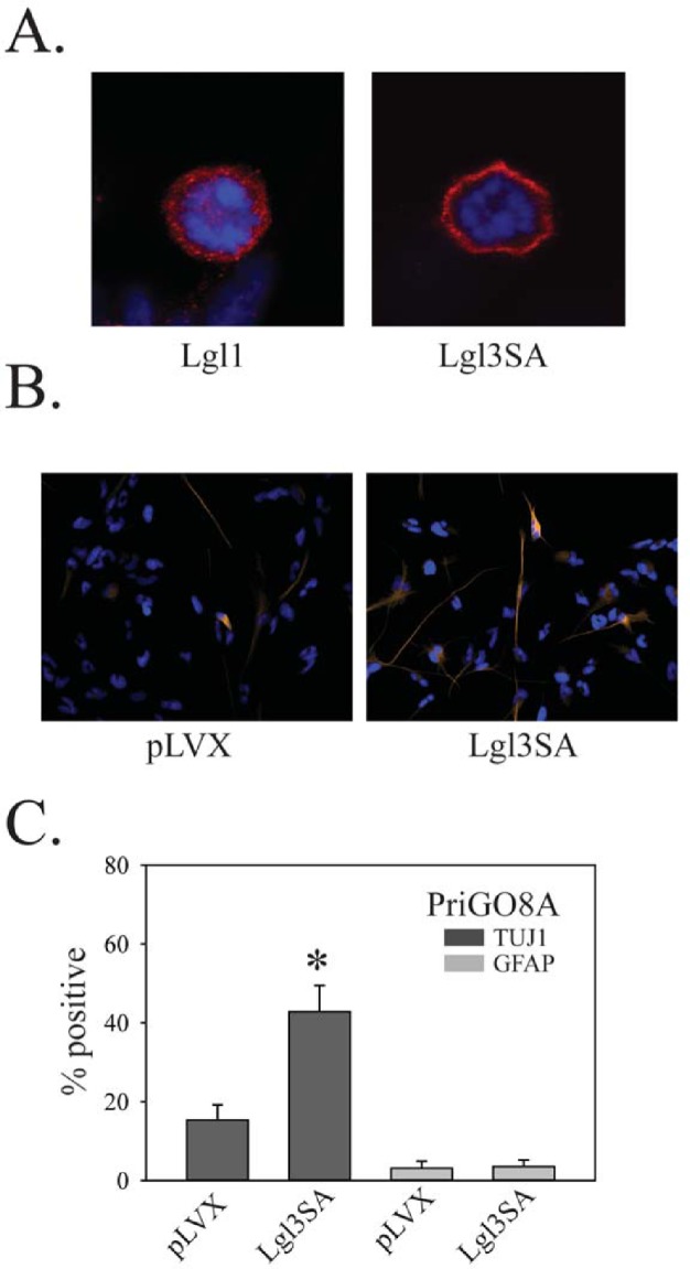
A. PriGO8A cells were transduced with either Lgl1 (left) or Lgl3SA (right). Four days later cells were fixed and immunofluorescence with anti-Flag antibody was performed. Representative examples of mitotic cells are shown. B. PriGO8A cells were transduced with either empty vector control (pLVX) or vector expressing Lgl3SA. Seven days later cells were fixed and immunofluorescence for TUJ1 and GFAP was performed. Representative images for TUJ1 staining are shown. C. Quantitation of TUJ1 and GFAP immunofluorescence seven days after transduction with Lgl3SA. Data are shown as the mean ± SE. * indicates a p value less than 0.05.