
Figure 2. Mitochondrial Activity Staining in Fresh Frozen Human Breast Cancer Tumor Tissue Sections.
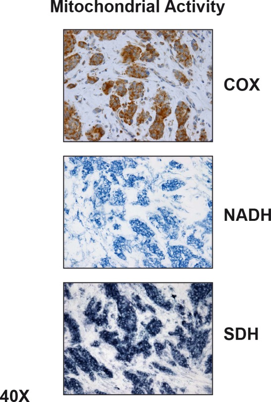
Note that epithelial cancer cell “nests” amplify their mitochondrial metabolism. In contrast, surrounding stromal fibroblasts show little or no functional mitochondrial staining, indicating that they show a shift towards glycolysis. COX, NADH, and SBH represent functional activity staining for mitochondrial complex IV, I, and III, respectively. Reproduced, with permission, from [58].