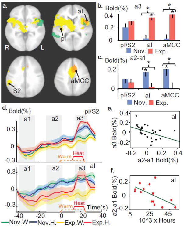
Figure 2. Expertise in OP modulates the temporal processing of painful stimuli.
a. Meditation experts had greater activity in primary pain regions during pain and decreased activity during the anticipatory period prior to pain. This is revealed by a voxel-wise group comparison of the response during (orange clusters) and prior to (green clusters) the pain stimulus (corrected, p<0.005). These maps are overlaid on the pain-related regions defined by the contrast heat vs. warm across groups (yellow clusters, t-test, corrected, p < 2×10^ −5). Talairach coordinates of axial views z= −2, 8, 31 and 42 mm. b. Experts differed more from novices in the anterior part of the pain-related regions than its sensory part during pain processing. The graph displays the response in sensory part of the pain-related regions, including posterior insula and secondary sensory cortex (labeled pI/S2), and in left aI (peak at (−38, 13, 7)) and aMCC (peak at (−8, 20, 39)) regions (in orange in 2a). Error bars are SEM. c. Experts had less anticipatory activity than novices in aI, aMCC but not in pI/S2. d. Average BOLD activity across time in pI/S2 and in left aI (baseline set to 0 at the onset of a1 for display purpose). e. Anticipatory activity in left aI predicted the pain-evoked response therein during pain controlling for Group factor and sensory activity in pI/S2 (Partial correlation r=−0.43, p<0.05). f. The amount of meditation practice in life for experts was negatively correlated to anticipatory activity in left aI (r= −0.63, p<0.05). * indicates p<0.05.

