
Fig. 4.
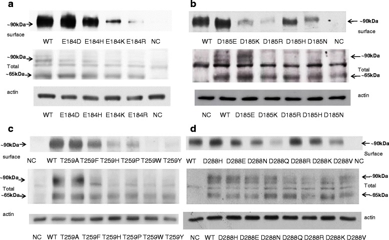
Protein expression of OATP1A2 and its mutants in HEK-293 cells. Western blot analysis of wild-type OATP1A2 and derivative mutants created at amino acid position 184 (a), 185 (b), 259 (c), and 288 (d). Upper panels, cell surface expression of OATP1A2 and its mutants. Cells were biotinylated, and the labeled cell surface proteins were precipitated with streptavidin beads and separated by gel electrophoresis, followed by Western blotting with anti-OATP1A2 antibody. Middle panels, Western analysis of total cellular expression of OATP1A2 and its mutants. Lower panels, after stripping, blots were reprobed with anti-actin antibody. WT, wild type; NC, negative control