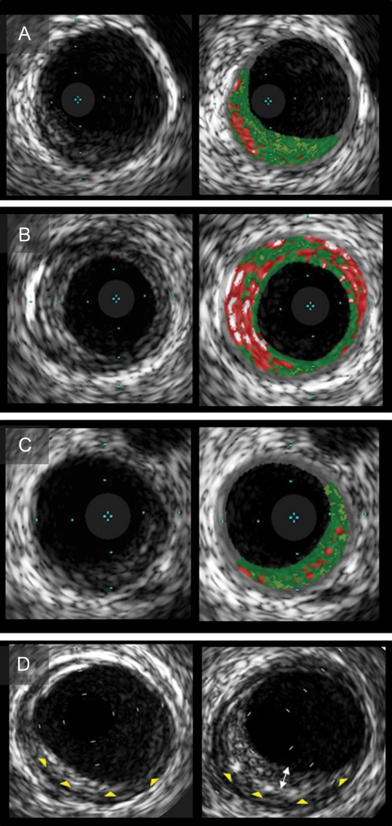
Figure 2.

Intravascular ultrasound images of plaques with multiple layers. Grayscale intravascular ultrasound demonstrated that multilayer appearance consisted of stratified layers with distinct echogenicity (A–C). Corresponding virtual histology-intravascular ultrasound images show a single (A), multiple layers of necrotic core (B), and plaque without necrotic core layer (C). (D) Multilayer formation during follow-up. A new layer (bidirectional arrow) was superimposed on the preexisting layer (arrow heads). Intravascular ultrasound indicates intravascular ultrasound.
