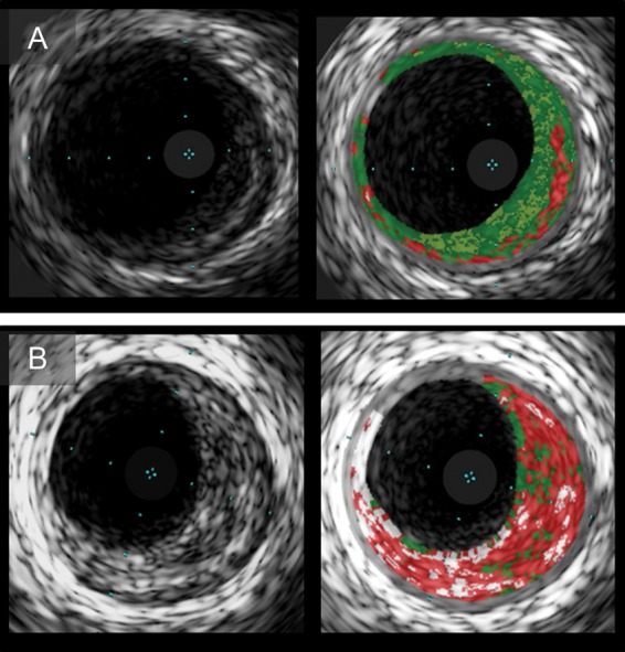
Figure 5.

Serial intravascular ultrasound images of plaques with multilayer appearance. Plaque with multilayer appearance at baseline (A) had subsequent multilayer formation at follow-up (B). Virtual histology-intravascular ultrasound images show a single layer of necrotic core (A) and multiple layers (B).
