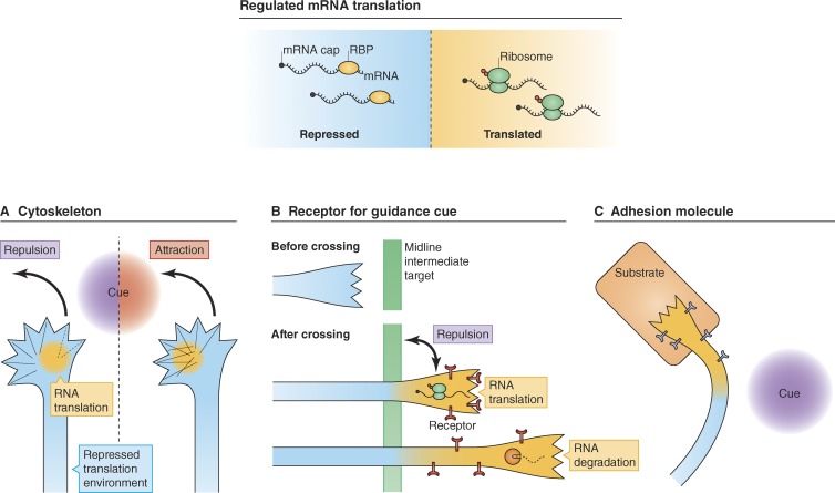
Figure 1.
How regulated mRNA translation mediates axon guidance. (A) Several studies support a model in which guidance cue–induced asymmetrical synthesis of cytoskeletal proteins, or their regulators, mediates attractive/repulsive responses in growth cones through the polarization of cytoskeletal dynamics (Wu et al., 2005; Leung et al., 2006; Piper et al., 2006; Lin and Holt, 2007). (B) During midline crossing of axons, the receptor Robo 3.2 for the repulsive guidance cue Slit present at midline intermediate targets is expressed only on distal axon segments after crossing. This spatiotemporal expression pattern is formed through translational control to avoid the repulsion between the midline intermediate targets and axons that have not yet reached the midline (Colak et al., 2013). The Robo3.2 transcript, which harbors a PTC, is degraded by the NMD pathway after local translation, modulating its expression levels (Colak et al., 2013). (C) Axonal translation of an adhesion molecule, NF-protocadherin, is triggered by regionally expressed Sema3A in the visual pathway, resulting in increased adhesion between axons and the substrate and helping axons to turn correctly in vivo (Leung et al., 2013). RBP, RNA-binding protein; Cue, guidance cue.

