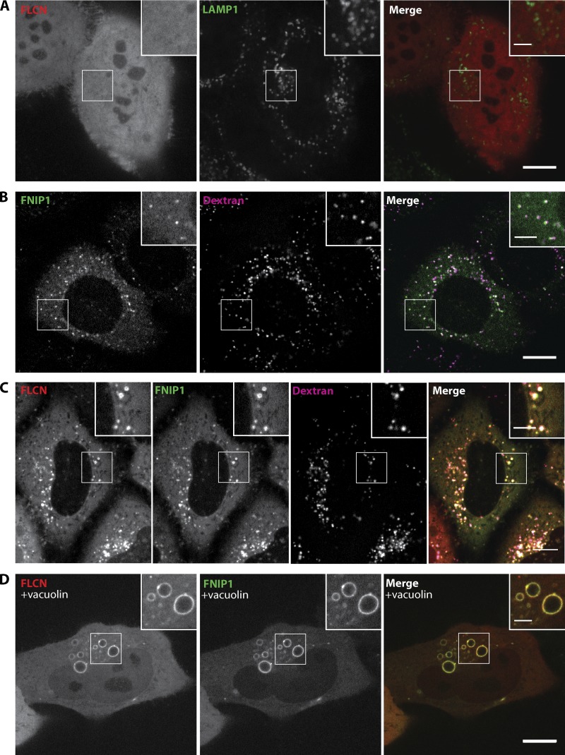
Figure 3.
FLCN and FNIP1 colocalize on lysosomes. (A) Localization of FLCN-tdTomato (red, diffuse) and LAMP1-mGFP (green, lysosomes). (B) FLCN-interacting protein 1 (FNIP1)-GFP (green) localizes to Alexa 647 dextran-positive (magenta) lysosomes. (C) Cells cotransfected with FLCN-tdTomato (red) and FNIP1-GFP (green) whose lysosomes were loaded with Alexa 647 dextran (magenta). (D) FLCN-tdTomato and FNIP1-GFP colocalization clearly occurs on the surface of the enlarged endosomes/lysosomes that form in vacuolin-treated (5 µM, 1 h) cells. All images were obtained by spinning disk confocal microscopy of live HeLa cells. Bars: (A–D) 10 µm; (insets) 5 µm.

