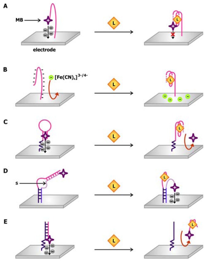
Figure 10.
Examples of electronic aptamer biosensors. a) A “signal-off” thrombin biosensor that uses methylene blue (MB) as a redox indicator [64]. b) A trans-signaling biosensor that depends on the interaction between [Fe(CN)6]3-/4-, a negatively-charged redox indicator, and the electrode [68]. c) A “signal off” thrombin sensor that exploits methylene blue's to function as both a DNA intercalating molecule and a redox indicator [69]. d) A structure-switching electronic aptamer biosensor in which the aptamer is bound by a redox dye-modified signaling oligonucleotide (S) in the absence of ligand [70]. Ligand binding displaces the oligonucleotide from the aptamer, allowing the dye to contact the electrode and generate a current. e) An electronic biosensor that signals by strand release [72].

