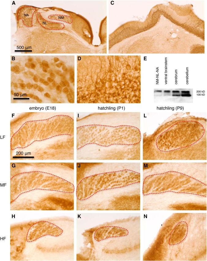
Figure 2.

mGluR II is expressed in NM neurons throughout the entire tonotopic axis across different ages. A, Immunohistochemistry revealed expression of mGluR II in auditory brainstem nuclei. NA, Nucleus angularis; NL, nucleus laminaris. B, Part of the NM shown at a higher amplification. C, D, Expression of mGluR II in the cerebellar cortex used as a positive control. The neuropils in the granular and Purkinje cell layers were intensely labeled, and the molecular layer was moderately labeled as a result of the extension of the dendrites of immunoreactive cells (primarily Golgi cells) in the granular layer. E, Western blot confirmed the specificity of the antibody against mGluR II in chicken brain tissues. The two immunoreactive products of 100 and 200 kDa correspond to monomeric and dimeric forms of mGluR2/3, respectively. F–N, Expression of mGluR II in different characteristic frequency regions (LF, MF, and HF indicate low, middle, and high frequency, respectively) in E18 (n = 2 animals; F–H), 1- or 2-d-old post-hatch chicks (P1–P2, n = 5 animals; I–K), and 1-week-old chicks (P7–P9, n = 2 animals; L–N).
