
Figure 6.
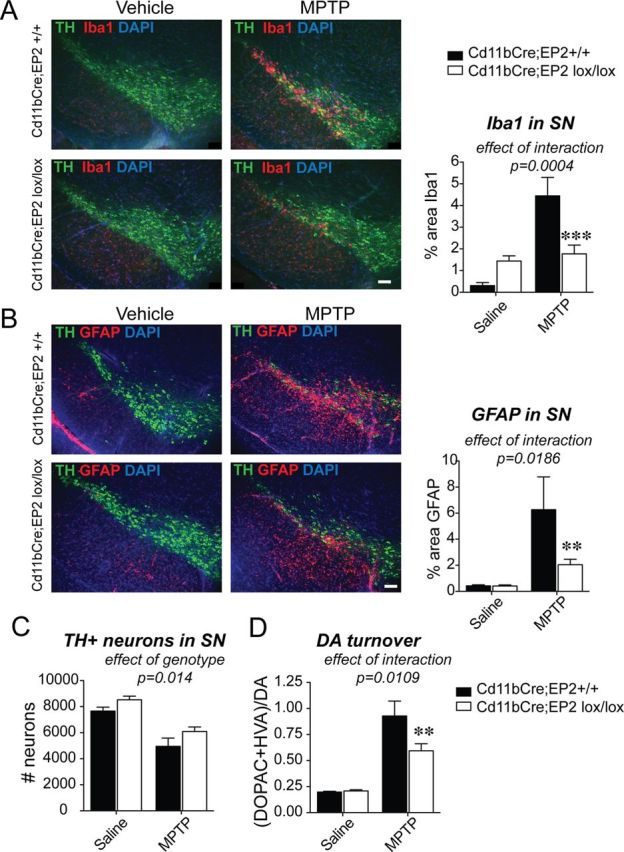
Conditional deletion of EP2 in microglia and macrophages reduces glial activation in the MPTP model of Parkinson's disease. Three-month-old male Cd11bCre;EP2lox/lox and control Cd11bCre;EP2+/+ mice were administered either vehicle or MPTP and SNpc was examined at 7 d after MPTP (n = 4–8 per group). A, Immunofluorescent staining of TH in dopaminergic neurons of the SN, Iba1 in microglia, and nuclear stain DAPI is shown for all four groups at 7 d after MPTP; (ANOVA effect of interaction, p = 0.004, Bonferroni post hoc ***p < 0.001; scale bar, 200 μm). B, GFAP immunofluorescence is significantly increased at 7 d after MPTP and is reduced in Cd11bCre;EP2lox/lox mice (ANOVA effect of interaction, p = 0.0186; Bonferroni post-test **p < 0.01; scale bar, 200 μm). C, Quantification of TH-positive neurons in SNpc shows effect of genotype and treatment only (p = 0.014 and p < 0.0001, respectively). D, DA turnover is significantly reduced with loss of microglial EP2 in MPTP-treated mice (ANOVA effect of interaction, p = 0.0109; Bonferroni post-test **p < 0.01).