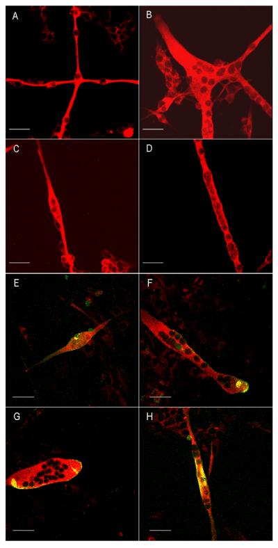
Figure 2.
Different morphologies of myotubes stained with embryonic, myosin heavy chain antibodies (Red) and clustering of acetylcholine receptors (Green) on the membrane surface of myotubes. Scale bar is 50 μm. A. Chain like morphology of the myotubes. B. Branched morphology of the myotubes. C. Spindle shaped morphology of a single myotube. B. Cylinder shaped morphology of a single myotube. E–H. Different morphologies of myotubes indicating the clustering of acetylcholine receptors (Green) on the membrane surface of different myotubes.

