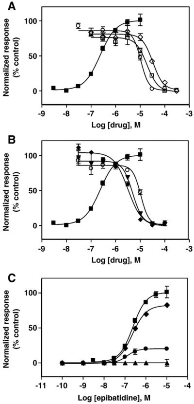
Fig. 1.
Effect of 18-methoxycoronaridine (18-MC), ibogaine, and PCP on (±)-epibatidine-induced calcium influx in TE671-hα1β1γδ cells. (A) Increased concentrations of (±)-epibatidine (■) activate the hα1β1γδ AChR with potency EC50 = 0.26 ±0.04μM (nH = 1.23 ±0.06). Subsequently, cells were pre-treated for 5min with several concentrations of 18-MC (○), ibogaine (□), and PCP (◊) followed by addition of 1 μM (±)-epibatidine. (B) Longer pre-treatment increases the inhibitory potency of 18-MC on (±)-epibatidine-induced calcium influx in TE671-hα1β1γδ cells. Pre-treatment periods were: 5 min (○), 4 h (▼), and 24 h (◆), respectively. Responses in both plots were normalized to the maximal (±)-epibatidine response which was set as 100%. The calculated IC50 and nH values are summarized in Table 1. (C) Pre-treatment with 1 (◆), 10 (●), and 100 μM 18-MC (▲), respectively, inhibits (±)-epibatidine-induced calcium influx in TE671-hα1β1γδ cells in a dose dependent and noncompetitive manner. The plots are representative of twenty-seven (■), nine (○), six (□), three (◊) and three (▲,▼,◆,●) determinations, respectively, where the error bars represent the standard deviation (S.D.) values.

