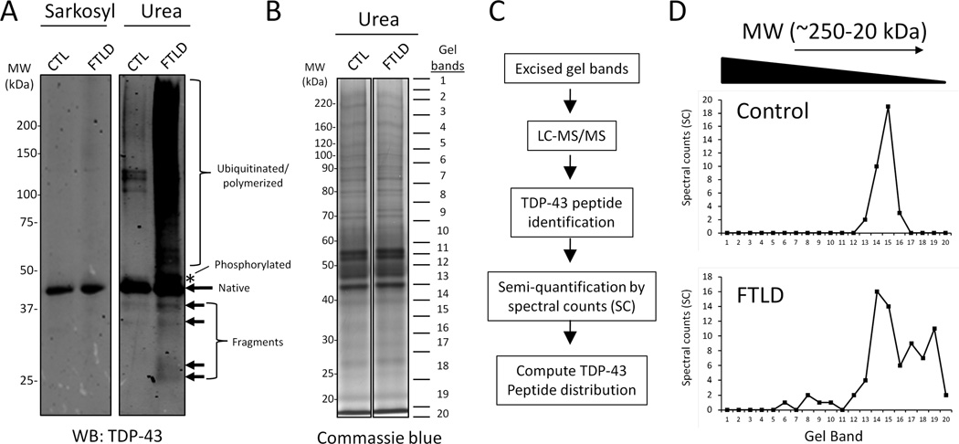
Figure 1. Identification of TDP-43 proteolytic fragments from post-mortem control and FTLD brain.
(A) Western blot analysis of sarkosyl (soluble) and insoluble fractions show enrichment of post-translationally modified TDP-43 (i.e. ubiquitination/polymerization, phosphorylation and proteolytic cleavage) in control and FTLD brain. (B and C) Detergent-resistant fractions were resolved by SDS-PAGE and the control and FTLD samples were excised into 20 gel slices, trypsin digested, and analyzed via LC-MS/MS on a high resolution Orbitrap mass spectrometer. (D) Differences in the TDP-43 peptide distribution between control and FTLD. Spectral counts (SCs) (x-axis) refer to the number of unique TDP-43 peptide sequencing events in each gel band (y-axis).

