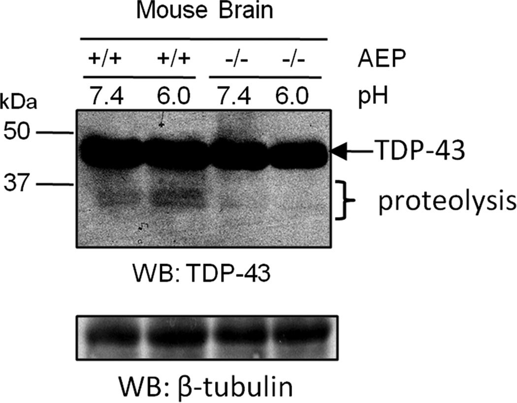
Figure 3. TDP-43 fragments are reduced in AEP−/− brain.
Comparison of whole brain homogenates from AEP-null (−/−) and wild-type (+/+) littermate controls by western blot revealed that N-terminal TDP-43-immunoreactive fragments (~35 and 32 kDa) were substantially reduced in the absence of AEP. β-tubulin was used as a western blot loading control. Data representative of two independent experiments.

